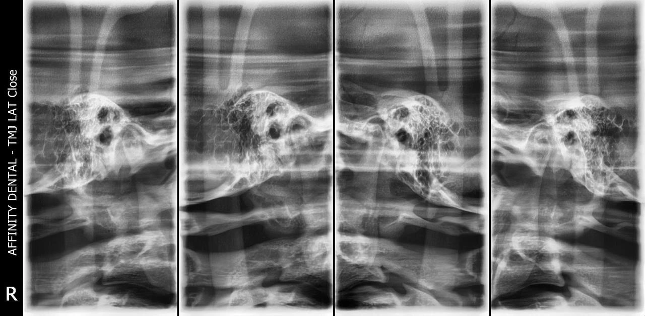
This set of transcranial X-ray images captures both the right and left temporomandibular joints (TMJ) in various states of movement—typically open and closed. It is used in diagnosing TMJ disorders, assessing condyle position, evaluating joint asymmetry, and monitoring structural changes in the joint over time. These views help dentists and specialists understand the functional condition of the jaw joints.